
Nasal Fibrolaryngoscope: The Diagnostic Standard Every ENT Specialist Must Master


Nasofibrolaryngoscopy is no longer a highly complex procedure reserved for large hospital centers. Today, it is the first-line diagnostic tool in any well-equipped ENT office. This article reviews its technical foundation, its clinical indications, and the criteria that define a quality device, designed for professionals who already use it or are evaluating incorporating it.
What is a nasofibrolaryngoscope and how does it work?
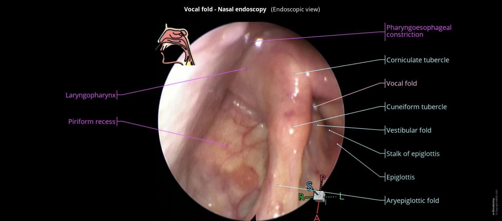
A nasofibrolaryngoscope is a small-caliber flexible endoscope that integrates an illumination system, an imaging camera, and, in some models, a working channel for biopsy or lavage. It is introduced through the patient's nasal cavity and allows real-time visualization of the entire upper airway: nasal cavities, choanae, nasopharynx, oropharynx, hypopharynx, larynx, and the upper third of the trachea.
Its operating principle is based on two main technologies that coexist in today's market. Fiber optic systems transmit the image through a bundle of glass fibers from the tip to the examiner's eye or the external camera. The more recent chip-tip systems place the image sensor directly at the distal end of the instrument, which eliminates the resolution losses inherent in fiber transmission and generates sharper images.
Functional anatomy that can be explored
- Nasal cavities: Mucosa, turbinates, septum, meatuses, and sinus drainage areas.
- Nasopharynx: Roof, torus tubarius, Eustachian tube orifice, and adenoids.
- Oropharynx and hypopharynx: Palatine tonsils, tongue base, piriform sinuses, and posterior wall.
- Larynx: Epiglottis, arytenoids, true and false vocal cords, subglottis.
- Proximal trachea: Upper third with assessment of tracheocartilaginous mobility.
Clinical indications: when to indicate nasofibrolaryngoscopy
The wide field of view of the nasofibrolaryngoscope makes it the most versatile diagnostic instrument in the ENT office. These are its main indications in Colombian clinical practice:
Laryngeal and vocal pathology
- Dysphonia of more than 2 weeks' duration
- Suspected vocal nodules, polyps, or cysts
- Vocal cord paralysis or paresis
- Reinke's edema
- Recurrent laryngeal papillomatosis
- Preoperative evaluation in laryngeal surgery
- Post-surgical vocal follow-up
Nasal, pharyngeal, and airway pathology
- Chronic nasal obstruction of undetermined cause
- Recurrent epistaxis without a visible focus
- Snoring and suspected OSAH
- Pharyngeal mass or asymmetry
- Dysphagia — FEES protocol
- Suspected neoplasm in the upper airway
- Difficult-to-locate foreign body
Persistent dysphonia of more than two weeks in an adult patient is a formal indication for nasofibrolaryngoscopy according to the guidelines of the American Academy of Otolaryngology. Failing to do so represents a diagnostic gap with medico-legal implications in the event of a late diagnosis of laryngeal carcinoma.
Fiber optic vs. chip-tip: differences that impact diagnosis
The choice of technological system is not just a budget decision. It has direct implications for diagnostic quality, patient comfort, and the profitability of the service in the medium term.
| Parameter | Fiber optic (FOE) | Chip-tip (digital) |
|---|---|---|
| Image resolution | Good. "Honeycomb" effect visible in small lesions | True HD or 4K. No loss of resolution |
| Distal diameter | 2.2 – 3.5 mm | 3.1 – 4.2 mm |
| Detection of subtle lesions | Limited in lesions smaller than 3 mm | Superior for dysplasia, early lesions |
| Endoscope lifespan | Variable. Fibers degrade with use | Greater. More robust digital sensor |
| Stroboscopy compatibility | Limited | Compatible with stroboscopy units |
| Acquisition cost | Lower initial investment | Higher initial investment |
| Total cost over 5 years | Frequent repairs increase the real cost | Lower total cost of ownership |
In what cases is fiber optic preferable?
Fiber optic systems remain a valid option for offices with a low volume of weekly endoscopic procedures, or for specialists who are incorporating the equipment for the first time and want to assess demand before a larger investment. They are also useful as backup equipment or for pediatric procedures where the ultra-thin diameter is decisive.
When does the investment in chip-tip justify itself?
When the volume exceeds 10 to 15 procedures per week, or when the office includes vocal pathology of medium or high complexity, the chip-tip system is clinically and economically justified. Early lesion detection, high-definition documentation for the medical record, and the ability to integrate stroboscopy are advantages that directly impact the quality of care.
Key technical specifications when evaluating equipment
Beyond the brand, there are objective parameters that define the clinical suitability of a nasofibrolaryngoscope. These are the ones that should appear on any technical data sheet that the specialist reviews before making a decision:
- Insertion diameter: Adults: 3.1 – 3.7 mm. Pediatric: 2.2 – 2.8 mm. Directly impacts patient tolerance.
- Field of view: Between 70° and 90°. A larger angle allows exploration of the hypopharynx without additional repositioning.
- Working channel: Present in larger diameter models. Necessary for guided biopsy, aspiration, or instillation.
- LED light source: longer lifespan and lower temperature. Xenon: more intensity. Both are valid depending on use.
Every active medical device marketed in Colombia must have a valid Invima sanitary registration. Its absence constitutes an irregularity before the MSPS and can compromise the habilitation of the service. Before any acquisition, verify the registration number on the official Invima portal.
Disinfection and maintenance protocol
The correct reprocessing of the nasofibrolaryngoscope is a clinical and legal responsibility. Flexible equipment is compatible with high-level disinfection (HLD) but not with steam autoclave sterilization, which would irreversibly damage the optical and electronic components.
Steps of the standard protocol between patients
- Preliminary cleaning: Remove residues with a damp gauze immediately after the procedure.
- Leak test: Verify integrity before immersing the equipment in a chemical solution.
- Enzymatic cleaning: Immersion in an enzymatic solution with mechanical action in the working channel.
- HLD: 2% glutaraldehyde or peracetic acid according to the manufacturer's times.
- Rinse and dry: With sterile or filtered water. Dry completely before storing.


