
Sigmoidoscopy: what it is, what it’s used for, and how it’s performed


Sigmoidoscopy: Procedure and Applications in Colon Diagnosis
In hospital and clinic gastroenterology departments, sigmoidoscopy is a diagnostic procedure that allows the final part of the large intestine to be evaluated using a minimally invasive technique. Although many people are more familiar with colonoscopy, sigmoidoscopy remains an important tool in diagnosing various digestive diseases.
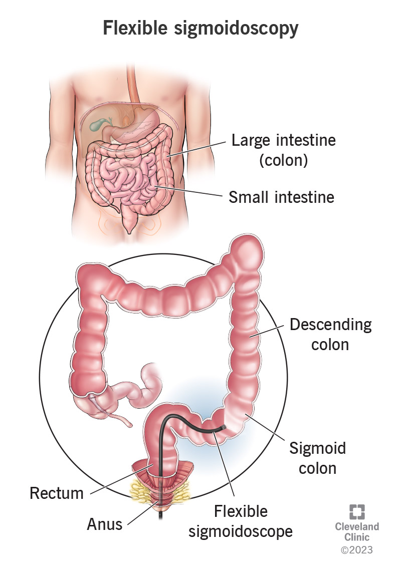
This procedure allows the rectum and sigmoid colon to be observed using a flexible endoscope equipped with a camera and lighting. Thanks to this technology, doctors can identify inflammation, polyps, bleeding, or lesions in this region of the colon.
In modern clinical practice, sigmoidoscopy is used for both the diagnosis and monitoring of certain intestinal diseases. It can also be a useful alternative in situations where it is not necessary to examine the entire colon.
Understanding how sigmoidoscopy works and when it is used helps clarify its role in digestive diagnosis.
What Is Sigmoidoscopy
Sigmoidoscopy is a medical procedure that allows the interior of the rectum and sigmoid colon to be examined using an instrument called a sigmoidoscope.
The sigmoidoscope is a flexible device that incorporates:
-
a high-resolution camera
-
an illumination system
-
channels for medical instruments
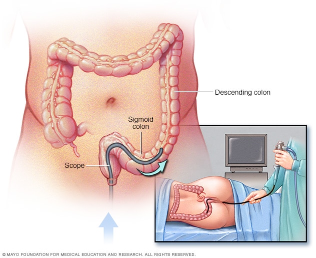
During the procedure, the instrument is inserted through the rectum and advanced toward the lower portion of the colon.
The images captured by the camera are transmitted in real time to a medical monitor, allowing the specialist to observe the intestinal mucosa in great detail.
This procedure allows for the detection of various abnormalities in the distal colon.
What Sigmoidoscopy Is Used For
Sigmoidoscopy is used to diagnose and evaluate several conditions of the large intestine.
Common clinical applications include:
-
evaluation of rectal bleeding
-
diagnosis of colon polyps
-
study of intestinal inflammation
-
diagnosis of ulcerative colitis
-
monitoring of inflammatory bowel diseases
It can also be used as part of early colorectal cancer screening programs in certain populations.
In many cases, sigmoidoscopy allows for the identification of lesions that can be treated during the same procedure.
Difference Between Sigmoidoscopy and Colonoscopy
One of the most frequent questions from patients is the difference between sigmoidoscopy and colonoscopy.
The main difference lies in the extent of the colon that is examined.
Sigmoidoscopy allows for the exploration of:
-
the rectum
-
the sigmoid colon
While colonoscopy allows for the examination of the entire colon.
Due to this difference, colonoscopy is usually used when a complete evaluation of the large intestine is required.
Sigmoidoscopy, on the other hand, may be sufficient when symptoms or clinical suspicion are localized to the final part of the colon.
Sigmoidoscopy Procedure in Clinical Practice
The sigmoidoscopy procedure is generally performed in endoscopy units within hospitals or specialized clinics.
The stages of the procedure usually include:
Bowel preparation
The patient may require a simple bowel preparation to clear the lower part of the colon.
This facilitates visualization during the examination.
Patient positioning
The patient is generally placed on their side on a medical stretcher.
Introduction of the sigmoidoscope
The doctor introduces the sigmoidoscope through the rectum and carefully advances it toward the sigmoid colon.
Visual examination
The images transmitted by the camera allow the specialist to examine the walls of the colon.
In some cases, biopsies can be performed or polyps removed during the procedure.
Technology used in sigmoidoscopy
The diagnostic quality of sigmoidoscopy depends largely on the technology used in the endoscopic system.
Modern systems include:
-
high-definition cameras
-
advanced light sources
-
digital image processors
-
video capture systems
These technological advances allow for the identification of small lesions that could go unnoticed with older equipment.
In hospitals that incorporate modern endoscopic technology, specialized providers such as GSMED Imaging participate in the implementation of these systems within gastroenterology departments.
Common errors when implementing sigmoidoscopy services
In hospital practice, some operational factors can affect the quality of the procedure.
One of the most frequent is inadequate bowel preparation, which makes it difficult to visualize the colon.
Problems can also arise when endoscopic system maintenance is not performed periodically.
Medical and technical staff training is essential to ensure safe procedures and reliable diagnoses.
Total cost of ownership of endoscopic systems
When hospitals evaluate implementing or upgrading technology for procedures such as sigmoidoscopy, they usually consider the total cost of ownership of the endoscopic system.
This analysis includes factors such as:
-
initial equipment investment
-
preventive maintenance
-
accessories and consumables
-
staff training
Evaluating these aspects allows for more informed decisions when acquiring medical technology.
A common case in digestive diagnosis
In a regional hospital seeking to improve the diagnosis of intestinal diseases, the medical team decided to expand the use of endoscopic procedures in patients with persistent digestive symptoms.
The implementation of sigmoidoscopy allowed for the rapid evaluation of the distal colon in patients with rectal bleeding or suspected colitis.
In many cases, the procedure allowed for the identification of early lesions without the need to perform more complex studies.
This type of experience demonstrates how sigmoidoscopy remains a useful tool in modern digestive diagnosis.


